Home » Without Label » Leg Anatomy Muscles Ligaments And Tendons : Expired website | This website has expired | Ankle anatomy ... / In the leg, muscle strains happen when a muscle is either stretched beyond its limits or forced into extreme contraction.
Leg Anatomy Muscles Ligaments And Tendons : Expired website | This website has expired | Ankle anatomy ... / In the leg, muscle strains happen when a muscle is either stretched beyond its limits or forced into extreme contraction.
Leg Anatomy Muscles Ligaments And Tendons : Expired website | This website has expired | Ankle anatomy ... / In the leg, muscle strains happen when a muscle is either stretched beyond its limits or forced into extreme contraction.. Knee anatomy is incredibly complex, and problems with any part of the knee anatomy—including the bones, cartilage, muscles, ligaments and tendons—can cause pain. Possibly the most important tendon in terms of mobility is the achilles tendon. Damage to the nervous system including the brain, spinal cord, and nerves degenerative diseases: Small tears of the tendon can make it difficult to walk and participate. A muscle strain is a stretch or tear of muscle fibers.
The tarsal bones are found near the. Damage to the nervous system including the brain, spinal cord, and nerves degenerative diseases: Your leg has several primary groups of muscle as well as several tendons. The calf muscle, on the back of the lower leg, is actually made up of two muscles: The hamstring muscles in the back of the thigh, the quadriceps muscles in the front, and the adductor (groin) muscles on the inside.
Ankle Anatomy - Orthopedic Surgery, Algonquin, IL ... from www.eorthopod.com Deltoid ligaments, which attach the tibia to the talus and calcaneus and provide stability to the insides of the ankles. Some of the more common ones are: Foot and ankle anatomy is quite complex. Tendons attach muscle to bone. The quadriceps and hamstring muscles work together to straighten (extend) and bend (flex) the leg. It provides the power necessary to straighten the knee. Your quadricep muscles, also known as quads, consist of four muscles that compose the front of your leg; Muscle and tendon pain in legs, muscles and tendons of the leg and foot.
The medial ligaments, sometimes called the deltoid ligaments, in the inner ankle the lateral ligaments, in the outer ankle both groups of ligaments help. Each of these muscles is a discrete organ constructed of skeletal muscle tissue, blood vessels, tendons, and nerves. Tibialis posterior tendon tibialis posterior is the deepest muscle on the back of the leg. The foot and leg muscle/tendon connectionto better understand foot and leg muscle/tendon injuries, it is important to appreciate the basic elements that enable your body parts to move. Anatomy leg muscles and tendons | online.kwc.edu author: A joint capsule is a watertight sac that surrounds a joint. Learn about the muscles, tendons, bones, and ligaments that comprise the knee joint anatomy. Deltoid ligaments, which attach the tibia to the talus and calcaneus and provide stability to the insides of the ankles. Damage to the nervous system including the brain, spinal cord, and nerves degenerative diseases: Included are several layered views of the back muscles, the dorsal muscles, subclavius muscles, rhomboideus major and minor muscles, deltoid muscles and many more. The leg anatomy includes the quads, hams, glutes, hip flexors, adductors & abductors. Ligaments, tendons, and muscles play an important role in the function of the hip. Clavicle fracture (broken collarbone) treatment.
There are many muscles located in the lower leg, but there are three that are particularly well known—the gastrocnemius and the soleus, which are the most powerful muscles in the lower leg, and the anterior tibialis. The knee joint is a complex structure that involves bones, tendons, ligaments, muscles, and other structures for normal function. The leg muscles are organized in 3 groups: A muscle strain is a stretch or tear of muscle fibers. Included are several layered views of the back muscles, the dorsal muscles, subclavius muscles, rhomboideus major and minor muscles, deltoid muscles and many more.
Ankle, Foot, and Lower Leg Ultrasound | Radiology Key from i1.wp.com The leg anatomy includes the quads, hams, glutes, hip flexors, adductors & abductors. Related posts of muscles and tendons of the leg muscle anatomy for gym. In the leg, muscle strains happen when a muscle is either stretched beyond its limits or forced into extreme contraction. The medial ligaments, sometimes called the deltoid ligaments, in the inner ankle the lateral ligaments, in the outer ankle both groups of ligaments help. Tendons vary in size and are somewhat elastic and attach bones to muscles. Ligaments connect two or more bones together and help stabilize joints. Deltoid ligaments, which attach the tibia to the talus and calcaneus and provide stability to the insides of the ankles. It provides the power necessary to straighten the knee.
When there is damage to one of the structures that surround the knee joint, this can lead to discomfort and disability.
Hip ligaments and tendons, tough. Some of the more common ones are: A muscle strain is a stretch or tear of muscle fibers. A number of tendons run through the ankle, attaching muscles of the lower leg to the bones of the foot and ankle. Posterior view of leg showing muscles and tendons involved in ankle movement. Bones, muscles, tendons, and ligaments in the leg muscles diagram above, there are many muscles that make up your legs and support it to move. Thigh muscle strain anatomy the thigh has three sets of strong muscles: Tibialis posterior tendon tibialis posterior is the deepest muscle on the back of the leg. The tarsal bones are found near the. Muscle anatomy for gym 12 photos of the muscle anatomy for gym muscle anatomy and fitness, muscle anatomy for fitness, muscle anatomy for gym, human muscles, muscle anatomy and fitness, muscle anatomy for fitness, muscle anatomy for gym In the leg, muscle strains happen when a muscle is either stretched beyond its limits or forced into extreme contraction. Because the leg has many different muscles, it is vulnerable to several different types of muscle strains. Ligaments connect two or more bones together and help stabilize joints.
Muscle anatomy for gym 12 photos of the muscle anatomy for gym muscle anatomy and fitness, muscle anatomy for fitness, muscle anatomy for gym, human muscles, muscle anatomy and fitness, muscle anatomy for fitness, muscle anatomy for gym One of the most important tendons is the quadriceps tendon. Related posts of muscle, tendons and ligaments of leg human human anatomy muscles abdominals. In the hip, the joint capsule is formed by a group of three strong ligaments that connect the femoral head to the acetabulum. It provides the power necessary to straighten the knee.
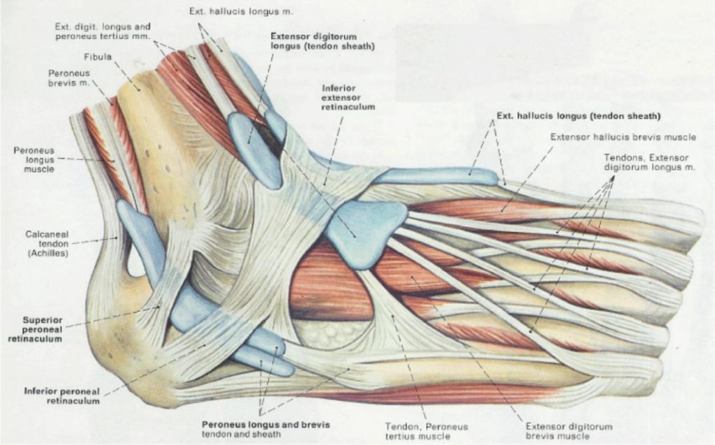
What is a Retinaculum? from corewalking.com Thigh muscle strain anatomy the thigh has three sets of strong muscles: A joint capsule is a watertight sac that surrounds a joint. One of the most important tendons in terms of mobility of the leg is the achilles tendon. Tendons attach muscle to bone. The tibialis posterior tendon is the main invertor of the foot and also helps the calf muscles to plantarflex the foot. To ensure your body moves smoothly with a minimum of friction, muscles are enveloped in a slippery skin like tissue called fascia. Tendons and ligaments are bands of connective tissue that help stabilize the body and allow movement. Collectively, the muscles in this area plantarflex and invert the foot.
Possibly the most important tendon in terms of mobility is the achilles tendon.
Because the leg has many different muscles, it is vulnerable to several different types of muscle strains. Foot and ankle anatomy is quite complex. The achilles tendon is also located in the lower leg. Small tears of the tendon can make it difficult to walk and participate. Clavicle fracture (broken collarbone) treatment. Collectively, the muscles in this area plantarflex and invert the foot. The hamstring muscles in the back of the thigh, the quadriceps muscles in the front, and the adductor (groin) muscles on the inside. Learn about the muscles, tendons, bones, and ligaments that comprise the knee joint anatomy. The quadriceps tendon works with the muscles in the front of your thigh to straighten your leg. Possibly the most important tendon in terms of mobility is the achilles tendon. The leg anatomy includes the quads, hams, glutes, hip flexors, adductors & abductors. The lower leg lies between the knee and the ankle. Muscles, either individually or in groups, are supported by fascia.